Diseases in livestock carry a special burden for commercial and small producers, practitioners, and the animals affected. While the most common causes of disease are viruses, bacteria, fungi, and parasites, prions and genetics can also come into play. Some of these disease have received a fair amount of attention over the years, others might surprise you with their symptoms, transmission, and sources.
As always, when faced with odd symptoms, collaboration with veterinarians is key and goes hand-in-hand with practicing solid biosecurity.
In the end, living organisms sometimes hold true to the old saying that, “If you’re going to have livestock, you’re going to have dead stock.” And, even though producers and veterinarians are working hard to figure out the what, why, and how we treat and avoid these diseases, many of them are still being studied.
Here are 10 weird animal diseases that we’ve come across in the livestock industry, and what to look for:
1. Hereditary Equine Regional Dermal Asthenia (HERDA)
This one’s just not even a little good (poco bueno). Hereditary equine regional dermal asthenia or HERDA is an inherited skin condition that’s primarily found in Quarter Horses and breeds with Quarter Horse lineage. Caused by a genetic mutation and typically attributed to the foundation sire Poco Bueno, HERDA was discovered in 1971. The disease is characterized by hyper-extensible skin that scars easily and leaves severe lesions, particularly along the back of affected horses.
According to UC Davis’s School of Veterinary Medicine, the mutation alters the proper formation of collagen, an integral structural protein in all connective tissues. In addition, other studies indicate that these horses may also suffer from corneal ulcers, decreased corneal thickness, increased corneal curvature, diameter, and mild corneal opacity, affecting the horse’s vision. Other studies indicate that affected animals may have weakened heart valves.
Although horses affected by HERDA are typically euthanized, a diagnostic DNA test is available to identify if horses are affected or if they are carriers of the mutation.
2. Equine protozoal myeloencephalitis (EPM)
Sorry opossums, it’s not you. It’s EPM. Equine protozoal myeloencephalitis, or EPM, is a disease in horses that affects the central nervous system — this means, the brain and spinal cord. Caused by the microbe Sacocystis neurona, EPM infection is typically attributed to opossums. Horses are infected when they come into contact with fecal matter in pasture or feed from the opossum host. Other animals such as armadillos, raccoons, skunks, and cats may host the microbe, but they can’t directly pass this disease onto horses.
Horses that are affected by EPM will show neurological signs such as stiff gaits or uncoordinated movements of lameness. Muscle atrophy may occur as well as drooping facial characteristics, difficulty swallowing, and loss of feeling. Horses may exhibit abnormal sweating, head tilting, and even be affected by seizures.
Several tests are available to diagnose EPM: Serum antibody tests, IgM capture ELISA blood tests, and spinal tapping. Infected horses may undergo a six-month antibiotic and anti-protozoal course, or a 28-day anti-protozoal course. While many horses will recover, early treatment of this disease is key. Controlling opossum populations and keeping feed covered will reduce the risk of infection.
3. Anthrax
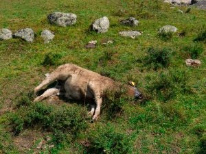
Glove up and get the flame thrower. Thankfully, anthrax infection is relatively rare in the United States. Caused by the Bacillus anthracis, a spore-forming bacterium, anthrax occurs in the soil. The disease is most common in herbivores, affecting domestic livestock, wildlife, and humans who may come in contact with the bacteria.
In cattle, sheep, and goats, staggering, trembling, difficulty breathing, convulsions, and death progress rapidly. Affected animals are commonly found dead, bloated, and with rigor mortis. Anthrax’s quick onset and course makes treatment difficult — in cases where live animals are diagnosed, prompt administration of penicillin is recommended by the AVMA.
Infections are controlled by removing livestock from the infected pastures, treating affected livestock, vaccinating unaffected stock, and burning carcasses. According tot he AVMA, necropsies should not be performed on animals that may have died from anthrax because of increased risk of exposure.
Anthrax affects humans in three forms: cutaneous, inhalation, and gastrointestinal. Anyone handling potentially infected carcasses should wear long sleeves, gloves, and a face mask.
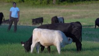
4. Testicular feminization
It’s a girl! Or, is it… Testicular feminization syndrome, or androgen insensitivity syndrome, is relatively rare, but when it’s diagnosed, it’s typically in a female-appearing animal that exhibits aggressive behavior typical of males in their species. When hormones are tested, these animals typically exhibit high testosterone levels. While external genitalia present as female, vaginal openings in these animals will end in a blind sac, with no cervix or uterus, and testicular tissue in place of ovaries.
In horses examined, chromosomal evaluations reveal that these animals have a male karyotype (X,Y). However, reproductive tissue’s insensitivity to androgens during development due to abnormalities in androgen receptors result in normal, external female genitalia. Removal of testicular tissue reportedly resolves aggressive behavior in these animals.
5. Meningeal worm infection
Got worms on the brain? Caused by the roundworm parasite Parelaphostrongylus tenuis, brain worms, deer worm, or meningeal worm infection causes neurological symptoms and death in goats, sheep, and camelids. Two words sum up treatment: difficult and expensive.
Areas with high rainfall and populations of whitetail deer may be at higher risk for infection. While deer seem to be able to pass Meningeal worms through their body with little effect, in domestic species, the parasite crosses the blood-brain barrier, migrating all over the body, causing intense itching, hind leg weakness, leg dragging, tremors, and the inability to stand. Pregnant animals are likely to abort their fetuses.
Treatment for meningeal worm infections includes high doses of fenbendazole, anti-inflammatory drugs to reduce the inflammation of nerve tissue. Early treatment is paramount in a successful outcome, because permanent spinal damage, including weak hindquarters and inability to deliver kids, may result from heavy infections. Animals that are unable to stand have a poor prognosis for recovery.
6. Equine odontoclastic tooth resorption and hypercementosis (EORTH)
Not so long in the tooth. Equine odontoclastic tooth resorption and hypercementosis or EORTH is a syndrome that typically occurs in older horses where the incisors and sometimes canine teeth’s roots are resorbed. As the roots are resorbed, the body attempts to stabilize the teeth by “cementing” bone-like substances to anchor the tooth, resulting in hypercementosis and swelling around the roots of affected teeth.
A slowly progressive disease, it’s unknown what triggers the process. Horses that are affected by EORTH may exhibit weight loss, attitude changes, salivation, head shaking, and other symptoms due to pain and discomfort. Radiographs are useful in making a diagnosis, and affected teeth are extracted.
7. Actinomyces
Lumpy jaw. Caused by the bacteria Actinomyces bovis, bony lump jaw is more common in cattle than in sheep and goats. Once a cut or abrasion is made by coarse feed or other objects, the bacteria enters the cut and migrates to the bone.
Once bone is infected, inflammation of the area may continue. The lower jaw is commonly affected. Bony masses under the mandible may be present with draining of pus-like fluid may ensue. The lymph nodes and throat may become enlarged, and animals may begin to salivate excessively, decreasing feed consumption due to pain.
Prognosis for affected animals is poor, and livestock with lumpy jaw are typically culled. Antibiotics, iodide administration, and removal of the affected bone and teeth can be removed in lieu of culling. Avoiding feeds that have abrasive seeds, or weeds in them can help avoid this issue.
8. Orf
Do you kiss your mother with that mouth? The scabby mouth virus also known as orf or contagious pustular dermatitis is a highly contagious disease that may impact people who come into direct contact with affected animals. The scabby lesions on the animals contain a live virus that’s easily spread, even though most infections clear up in four to six weeks.
Although it typically occurs in younger livestock, ewes without immunity may be affected when lambs suckle their teats, leading to ewes refusing to feed their lambs. Vaccines are available against orf, and lesions may be treated with antiseptics.
Orf vaccines are available, but typically recommended only when there is an outbreak. Lower stocking rates, raising a closed herd, and lambing outside are recommended to reduce and control orf transmission. It is also recommended that shed scabs and lesions are burned and buildings are disinfected with iodophor or formaldehyde.
9. Grass tetany
Not enough of a good thing. Winter and spring is the time of year to look out for signs of grass tetany or milk fever, a fatal metabolic disease. Associated with low levels of magnesium in livestock blood, animals grazing cool season grasses while lactating are at particular risk, though other growing stages of animals may be affected.
While livestock can hold magnesium in their bones and muscles, they cannot readily access these reserves. Animals suffering from grass tetany are often found dead following convulsions and a coma. Early signs include excitability, muscle twitching, stiff gaits, aggression, and staggering.
Prevention is key in this disease. Forage and soil testing, supplementing magnesium, and ensuring that quality feed are present. Affected cattle may be treated by restoring blood magnesium levels through intravenous calcium and magnesium.
10. Hemorrhagic bowel syndrome
We don’t know why. Hemorrhagic bowel syndrome occurs with a sudden onset of abdominal pain in dairy cattle, progressing to downed animals, shock, and finally, death. Although we’re not quite sure why HBS occurs, it’s a syndrome that’s occurring more frequently in mature, lactating dairy cattle and sometimes, in beef cattle.
Scientists and veterinarians believe that HBS is multifactorial. Risk factors for HBS may include a high amount of fermentable carbohydrate in the diet, dry matter intake, level of milk production, lactation number, herd size, season, and the presence of the causative organism in the feedstuff. Clostridium perfringens type A, a naturally-occurring inhabitant of cattle’s digestive tract may be one of the culprits. Another agent may be Aspergillus fumigatus, a fungus found in feed and forages.
In clinical findings, cattle affected by HBS have a history of anorexia, depression, drops in milk production, abdominal distention and pain, dark feces, and become dull and weak. Most cattle affected by the syndrome die within days even with fluid and electrolyte therapy.
Heidi Crnkovic, is the Associate Editor for AGDAILY. She is a New Mexico native with deep-seated roots in the Southwest and a passion for all things agriculture.